The birth of consciousness: a story of brain creation.
Human creation? Most of us are familiar with the biblical story of Adam and Eve. For those less religiously inclined, reproduction might spring to mind. The origin of our inception has long burdened humankind. How about an alternative philosophy? The intention of which is not to replace the Book of Genesis or contradict scientific truths, but to create a ‘brainy’ insight about the beginning. Let’s consider the profound René Descartes quote ‘I think therefore I am’ (Fig. 1).
If conscious thought is what makes us human, then brain creation is intrinsically linked to human creation.

Figure 1: Rene Descartes: I think therefore I am.
Have you ever wondered how your brain is created?
How the most complex entity in existence comes to be? The answer emanates from the microscopic, but miraculous, embryo. Succeeding the unlikely event of a sperm reaching and fusing with an egg, the fertilised egg splits into two identical copies of itself, which then continue to multiply in number to form a ball of cells. This cell cluster is the embryo, and these cells, called ‘stem cells’, have the ability to become any human cell type (e.g skin, blood, bone etc.). At first the embryo stem cells simply multiply in number, but once the embryo establishes its home inside the mother’s womb, these cells are ‘forced to choose’ one of three fundamental cell fates: the ecto-(outer), meso-(middle) or endo-(inner) derm (cell layer) (Fig. 2).
The three-week-old, ‘pea-sized’ embryo consists solely of these three cell layers.
The outer ectoderm layer eventually becomes outer-body parts (e.g. skin, hair and teeth), the middle mesoderm layer develops into bones, muscles and blood vessels, while the inner endoderm layer forms our inner-body compartments (e.g. gut and lungs).
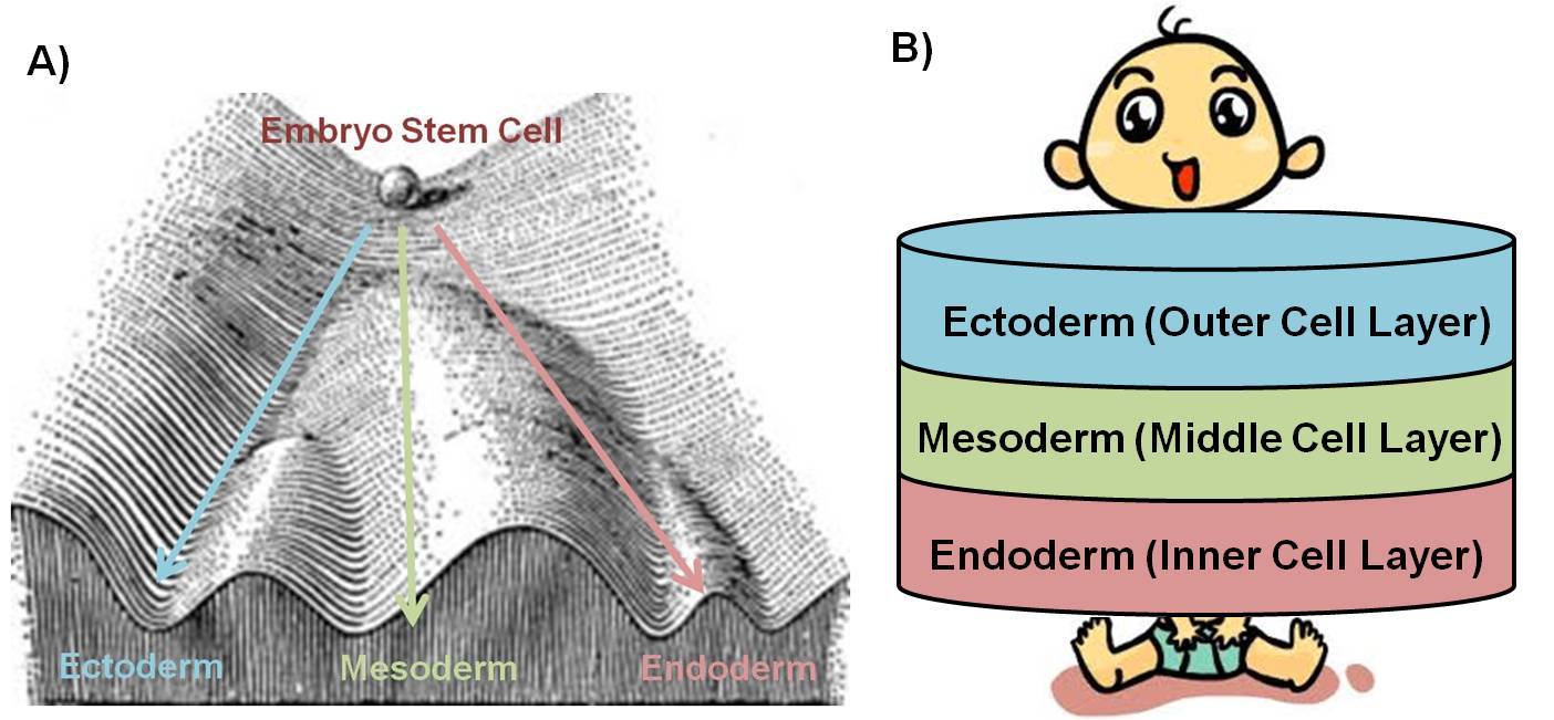
Figure 2: Embryo stem cell becomes ectoderm, mesoderm or endoderm.
So where does our brain come from? To state that embryo stem cells are ‘deciding’ their fate is not entirely accurate. In fact, the surrounding environment largely dictates what these stem cells become. It seems that peer-pressure even occurs in the womb, as neighbouring cells send ‘chemical’ messages to nearby stem cells to instruct their destiny.
Our brain arises as a result of such instructive interactions between cells in the developing embryo.
To spare us the scientific jargon, brain creation in the embryo will be compared to the ‘creation of brain-iness’ in school. In this curious analogy, a special mesoderm ‘rod’, which forms at the centre of the mesoderm (Fig. 3), acts as the teacher, while the overlying ectoderm cells are the unfortunate students. The mesoderm rod sends a suitably named chemical message called ‘Noggin’ towards the ectoderm, very much like how a teacher commands students to ‘use their noggin’ in school. The students that choose to listen to their teacher’s instruction, usually sitting eagerly right in front of the teacher, become ‘brainy nerds’.
In a similar fashion in the embryo, the ectoderm cells which lie directly above the mesoderm rod respond to the Noggin, sent by the rod, and become brain cells.
The ectoderm cells that do not respond to the message from the mesoderm rod, because of their distance away from the rod, continue as ectoderm cells which then become skin cells. Just like those stubborn students who sit far away from their teacher and ignore their instructions, continue unaware on their path to becoming ‘skin-heads’. Please excuse the excessive stereotyping. ‘But what about those impartial students who get caught between the brainy nerds and the skinheads’, you most certainly are not asking yourself? Well there is an important type of cell that fits this contrived metaphoric description too.
The cells that sit at the border between the ectoderm cells and the brain cells, marginally respond to the Noggin released by the mesoderm rod to become ‘neural crest’ cells (named so because of their position at the ‘crest’ of the brain/‘neural’ cells).
These neural crest cells travel to different parts of our body to become ‘peripheral’ brain cells, and have a variety of functions depending on where they end up. For example, the neural crest cells which travel to our heart settle there to control its beating, while those that get stuck in the gut control our digestion. The neural crest cells really only fulfil their destinies once they find their homes in our body. In the final indulgence of school-related analogies, neural crest cells could be compared to those indifferent students that travel around the globe before finding a suitable niche and purpose.
At this point of our embryonic journey, brain cells have arisen, however this is just the beginning.
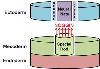
Figure 3: Neural plate (brain stem cells) and neural crest form from the ectoderm in response to noggin released by underlying rod of mesoderm.
These cells are in fact brain ‘stem’ cells, capable of becoming any type of brain cell, and have a long way ahead before they create our brain. The brain stem cells, which have emerged in the centre of the ectoderm, are collectively known as the ‘neural plate’. Over a short period of time, this neural plate expands, curls-up and then folds-in on itself to form the ‘neural tube’ (Fig. 4).
The neural tube, which runs from head to tail(-bone) at the centre of the embryo’s back, is the precursor of our brain and spinal cord.
When the neural tube forms it literally ‘gets under our skin’, by detaching from the ectoderm, and then positioning itself below the ectoderm and above the mesoderm.
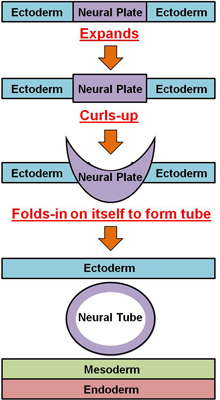
Figure 4: Neural plate expands, curls-up and then folds-in on itself to form the neural tube, which then lies under the ectoderm and above mesoderm.
Remember the mesoderm? The ‘middle’ stem cell layer that forms muscles? Let’s embark on another round of analogous stereotyping and imagine the mesoderm as one of those ‘muscular’ body-builders who use their force to make others succumb to their will.
This ‘muscular’ mesoderm ‘forces’ the brain stem cells in the front neural tube region to turn into ‘motor’ brain cells, which trigger our muscles movements (Fig. 5).
The ‘muscular’ mesoderm achieves this by sending a chemical messenger aptly named ‘sonic hedgehog’ (in honour of the blue, spiky-haired and fast-moving videogame character). Forgive the digression, but biology very rarely lends itself to such amusing nomenclature. Overlying the ‘backside’ of the neural tube is the ectoderm, the ‘outside’ stem cell layer that becomes skin. Because the ectoderm/skin is in contact with the environment, let’s think of the ectoderm as a ‘sensitive environmentalist’ who graciously attempts to recruit others to join their cause.
At the same time that the motor brain cells are generated, the 'environmental’ ectoderm ‘sensitively asks’ the nearby stem cells in the back neural tube region to become ‘sensory’ brain cells, which sense touch, pain and position from our environment.
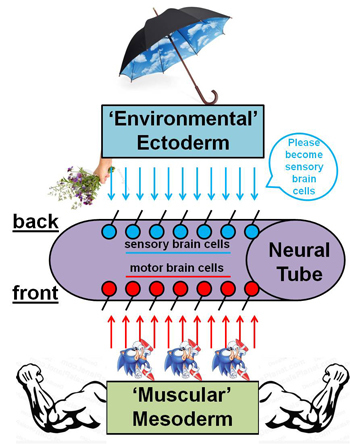
Figure 5: Mesoderm instructs front neural tube brain stem cells to become motor brain cells, while the ectoderm instructs back neural tube brain stem cells to become sensory brain cells.
How does our exceptional brain grow from this relatively uniform tube of brain stem cells? Well, unique signals at the embryos’ head region instruct the exponential growth of the top-end of the neural tube generating a large ‘swelling’/vesicle, which will eventually become the brain (Fig. 6). The continuous swelling of this developing brain, due to multiplication and growth of brain cells, results in the splitting of the brain vesicle into two ‘cerebral (brain) hemispheres (half-spheres)’. As it expands, the weight of the growing brain causes the neural tube underneath to bend, and the brain to fall-over and sit perpendicularly/‘at a right angle’ to the top of the spinal cord.
By birth, the brain has grown so large that it is forced by the skull to fold-in on itself, giving the brain its ‘ridgey’ appearance.
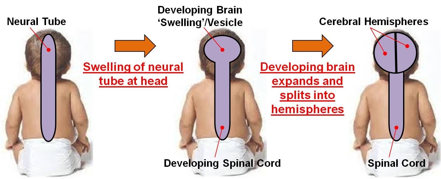
Figure 6: Unique signals instruct swelling of neural tube at head, and the developing brain then expands and splits into two cerebral hemispheres.
The disproportionate growth of the human brain during evolution (over the past millions of years), means that our brains are 5 times the size you would expect for a mammal our size. The unique head signals also direct the generation of a variety of brain parts/‘lobes’, including the frontal, parietal, temporal and occipital lobes, each with a generalised function (Fig. 7a). For example, the occipital lobe is where we produce sight (from visual images gathered by the eyes).
The most important lobe in terms of what ‘makes us human’ is the frontal lobe, as it carries out our higher mental processes such as thinking, decision making, and planning.
Within the different lobes of the brain, there are a number of brain regions which have specific roles. For example, the ‘hippocampus’ region is where we form our memories (perhaps the saying should read ‘memory of a hippo’ rather than an elephant). To make things even more confusing, throughout the brain different populations of brain cells are formed, which work together to carry out their particular task. For example, the ‘mid-brain cell population’ help us to control how, when and if we move. The death of these cells causes Parkinson’s disease (see https://svh_neuro.svbtle.com/parkinsons-disease-can-we-move-in-the-right-direction), which means that the sufferers cannot move in the way that they want (symptoms: slow, stiff and no movement), or stop ‘unwanted’ movements (symptoms: ‘shakes’ and tremors).
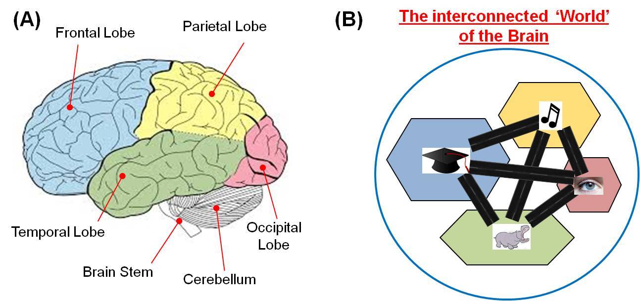
Figure 7: The different brain lobes, regions and populations interconnect via nerve fibers.
To put these subdivisions of our brain into perspective, imagine our brain as the world, the ‘lobes’ as its continents, the brain regions as countries and the individual brain cell populations as cities (Fig. 7b). These brain cell populations do not remain isolated, as each brain cell grows/‘sends-out’ nerve fibers/‘wires’ which connect with a number of other populations. Such connections can be visualised as motorways between major cities. Depending on the chemical signals that brain cells release, known as ‘neurotransmitters’, brain cells either excite (send messages via electrical impulses) or inhibit (stop these messages being sent) the cells they communicate with.
As a global community, these different populations of brain cells constantly work together to perform three major roles: 1) To receive and interpret information from our body, e.g. ‘ouch the boiling kettle is burning my hand’; 2) To send information around our body, e.g. ‘move my hand away from the boiling kettle’; and 3) To store information as memories, e.g. ‘do not touch a boiling kettle’.
The crowning part of our brain, called the ‘cerebral cortex’, is responsible for the supreme levels of conscious thought in humans. The exaggerated size of the human cerebral cortex sets us apart from other animals, and is the outstanding achievement of human evolution.
Buried beneath our enormous cerebral cortex are the ‘animalistic’ parts of our brain, which are unconscious and function in emotion, motivation, memory and sleep.
Between the brain and spinal cord is the ‘brain stem’, where the populations of brain cells that gift us our five special senses, smell, vision, taste, hearing and balance, are formed. Sitting on the back of the brain stem is the ‘cerebellum’ (meaning ‘little brain’), and looks like the brain’s ‘mini-me’. The cerebellum ensures the brain doesn’t make mistakes when controlling our body’s complicated movements (e.g. running, jumping or kicking), to give us balance and coordination. In fact, ‘Punch-Drunk Syndrome’ is a condition that affects boxers and alcoholics in which damage to the cerebellum causes a loss of balance and coordination (it is possible to temporarily self-inflict such symptoms following a session of alcohol consumption).
Below the brain stem, the spinal cord is responsible for the movement and sensation of our body.
As we know from above (how could we forget sonic hedgehog?), the front part of the spinal cord contains motor brain cells and the back part of the spinal cord contains the sensory brain cells. These motor and sensory brain cells project nerve fibres out of the spinal cord, which find their way to every corner of our developing bodies by following the routes laid-down by blood vessels.
The creation of reciprocal/‘two-way’ connections (via nerve fibres) between the brain, brain stem, cerebellum and spinal cord allows the brain to control, and interpret information from the entire central nervous system, and therefore our body. These connections form in such a way that our entire body is mapped out along the top of our cortex, right along its centre.
The brain’s body map is called the ‘homunculus’, meaning 'little man’.
This map is quite peculiar because our body is upside-down (our feet are at the top and our heads are at the bottom) and disproportionate (our hands and face get a larger part of the map than our limbs - these body parts are more sensitive and need more movement control, meaning they require more brain cells) (Fig. 8). What is even stranger about the way our brain connects with our body is that the right side of the brain controls the left side of our body, and vice versa.
The evolutionary or developmental advantage of having ‘crossed-wires’ (nerve fibres going from one side of brain to the opposite side of the body) remains a mystery.
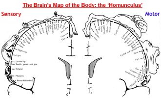
Figure 8: The brain’s map of the body, known as the Homonculus.
At birth, almost all the brain stem cells have turned into their final, life-long cell types, and have made connections with brain cells in the same and different parts of the brain. Astonishingly, our brain consists of at least several hundred distinct types of brain cells. Throughout life, the continuous establishment, alteration and refinement of brain cell interconnections facilitates the complex functioning of our brain.
A clear example of this strengthening and weakening of brain connections during adult life can be seen in the formation (learning), and subsequent loss (if not reinforced/recalled), of a memory.
Communication between the brain and the different parts of our nervous system is vital for our bodies to function normally throughout life. The most dramatic consequence of broken communication between the brain and our body can be seen in spinal cord injury (Fig. 9). For example, damaging the spinal cord at the neck means the brain loses control over, and sensation from, the body below the neck which results in quadriplegia.
What makes injuries that sever brain connections even more devastating is the unfortunate fact the brain cells lose the ability to grow nerve fibres around the body after we are born.
The special signals present during our development that instruct brain cells to project nerve fibres around our bodies are absent once we are born, meaning our bodies do not repair nerve connections that are severed.
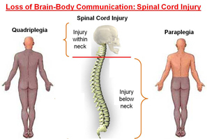
Figure 9: Loss of brain-body communication caused by spinal cord injury.
The precision at which the marvellous community of ~100 billion brain cells is formed is no less than a miracle. Its biological brilliance reflects millions of years of evolutionary adaptations and reconstructions, as well as masterful developmental processes. For example, during brain development an excessive number of brain cells are generated initially.
In an extraordinary process of refinement, the brain cells then compete for survival subject to the body’s requirements which ensures that only the correct number of brain cells remain.
Over the last few decades neuroscientists have begun to understand how interconnected brain cells communicate with one another to allow us to perform complicated tasks. Indeed, Professors John O’Keefe and Edvard and May-Britt Moser were awarded the 2014 Noble Prize for showing how brain cells arrange and communicate to give us our ‘inner GPS’. Recently introduced technologies will hopefully allow us to increase our understanding of the complex brain in the future. For example, the new ‘optogenetics’ approach allows us to see neurons when they are active (they literally light up).
Human nature and consciousness arise from the brain. Concomitantly, the brain is responsible for our experience of, and acts as the interface between, the self and the outside world. It constructs our memories of the past and our visions of the future.
Everything we think, feel, remember and dream is written by these brain cells.
It is almost incomprehensible how amazing our brains really are. But thanks to our brain’s intellect, we can aspire to appreciate its magnificence. By understanding the creation of our brain, we are a little closer to understanding what makes us human.
A version of this article appeared on the PLOSNeuro website as part of their #BAW2016 campaign to celebrate Brain Awareness Week 2016. See this version of the article below:
http://blogs.plos.org/neuro/2016/03/14/baw2016-the-birth-of-consciousness-a-story-of-brain-creation-by-shane-hegarty/